
Blog
260 and 340: The Two Peaks That Tell NAD+ From NADH

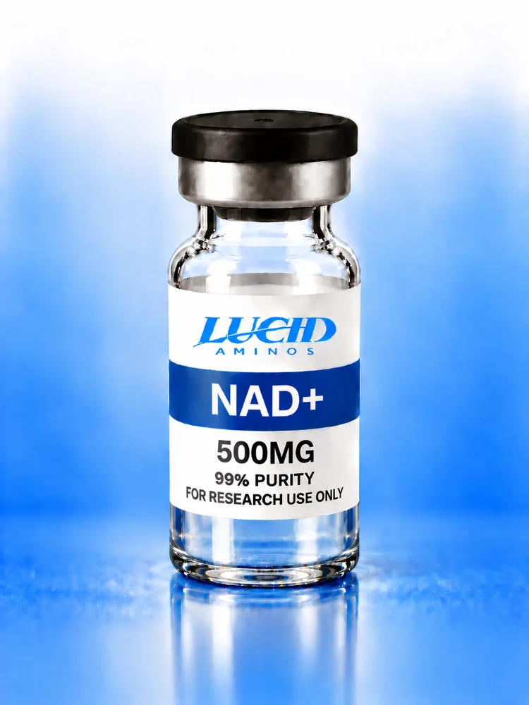
Place NAD+ in a spectrophotometer and it shows a strong peak at 260 nm. Place its reduced partner NADH in the same instrument and a second, smaller peak appears at 340 nm. That tiny shift made modern enzymology possible.
Where the 260 peak comes from
The adenine ring absorbs at 260 nm in both forms. The molar absorptivity is roughly 18,000 M⁻¹cm⁻¹, so a 0.1 mM solution in a 1 cm cuvette reads near 1.8. That is a quick way to estimate concentration.
Where the 340 peak comes from
When the nicotinamide ring picks up a hydride and becomes NADH, it gains an absorption band at 340 nm with an absorptivity of about 6,220 M⁻¹cm⁻¹. NAD+ has essentially none there.
Why that is useful
If an enzyme reaction converts NAD+ to NADH, you can watch absorbance at 340 nm climb in real time. The slope is the reaction rate. Otto Warburg’s lab used this principle in the 1930s, and it remains a staple of undergraduate labs.
A worked example
A rise of 0.311 at 340 nm in a 1 cm cell means 0.05 mM of NADH was produced (0.311 ÷ 6,220 = 0.00005 M). Keep reading and you will see why that simple division is so powerful.
For the research compound itself, see NAD+ 500mg.
Explore Lucid Aminos
- Shop all research compounds
- Read the lab reports (COA)
- Frequently asked questions
- Partner program
- About Lucid Aminos
- Contact us
For laboratory research use only. Not for human or veterinary use, and not intended to diagnose, treat, cure or prevent any condition. This article is educational and is not medical advice.