
Blog
Circular Dichroism: How Researchers Read the Shape of a Peptide

You cannot look at a peptide under a microscope and see its shape. But you can shine a special kind of light through it and read the answer from how the light changes.
The idea
Circular dichroism (CD) uses light that spirals left or right. Chiral molecules, such as the L-amino acids in peptides, absorb the two spirals by slightly different amounts. The difference is the CD signal.
Reading the curve
| Shape | Typical far-UV signature |
|---|---|
| Alpha helix | Two dips near 208 and 222 nm |
| Beta sheet | One dip near 218 nm |
| Random coil | A strong dip near 198 nm |
Why chemists like it
It needs only a small amount of sample and takes minutes. It can show whether a peptide folds when conditions change, for example when a helix-promoting solvent such as trifluoroethanol is added.
Limits
- It gives an average, not an atomic picture.
- Buffers that absorb UV can ruin the signal.
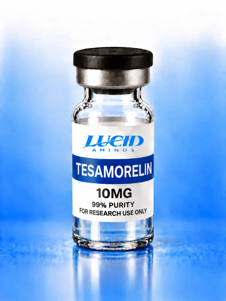
Long peptides such as Tesamorelin 10mg are the kind that CD can tell something about, while very short ones are mostly random coil. Keep reading.
Explore Lucid Aminos
- Shop all research compounds
- Read the lab reports (COA)
- Frequently asked questions
- Partner program
- About Lucid Aminos
- Contact us
For laboratory research use only. Not for human or veterinary use, and not intended to diagnose, treat, cure or prevent any condition. This article is educational and is not medical advice.